
Australia is a beautiful country and experiences some amazon weather in different states. Australia is known for its beautiful beaches, warm climate, and outdoor lifestyle. However, it is also known for having one of the highest rates of skin cancer in the world. In fact, skin cancer is the most common cancer in Australia. As per Sunsmart, More than two in three Australians will be diagnosed with skin cancer in their lifetime.
This is a significant health concern, and the question is: why does Australia have such a high rate of skin cancer?
UV Radiation
The answer to this question is multifaceted, and there are several contributing factors to consider. The first and perhaps most obvious reason is the country’s location. Australia is located near the equator, which means that the sun’s UV rays are stronger and more intense than in other parts of the world. This increased exposure to UV radiation means that Australians are at a higher risk of developing skin cancer.
UV radiation is classified into three types based on wavelength:
- UVA (315-400 nm): UVA rays have the longest wavelengths and can penetrate deep into the skin. They are primarily responsible for skin aging and can contribute to skin cancer.
- UVB (280-315 nm): UVB rays have shorter wavelengths and are more energetic. They are the main cause of sunburn and play a significant role in the development of skin cancer.
- UVC (100-280 nm): UVC rays are the most dangerous, but they are mostly absorbed by the Earth’s ozone layer and do not reach the surface.
The Impact of UV Radiation on Skin Health
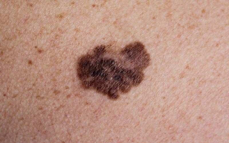
UV radiation can cause significant damage to the skin, leading to various types of skin cancer. The primary effects include:
DNA Damage
UV radiation can directly damage the DNA in skin cells. This damage can cause mutations, which may lead to the uncontrolled growth of cells, resulting in skin cancer. The body attempts to repair this damage, but repeated exposure over time can overwhelm the repair mechanisms, increasing the risk of cancer.
Immunosuppression
UV radiation can suppress the local immune response in the skin, making it easier for cancerous cells to develop and proliferate. This immunosuppression can also reduce the skin’s ability to fight off infections and other skin conditions.
Outdoor Lifestyle
Another contributing factor is the country’s outdoor lifestyle. Australians love to spend time outdoors, whether it’s at the beach, playing sports, or simply enjoying the beautiful scenery. This means that people are often exposed to the sun for long periods of time, which can increase their risk of developing skin cancer.
In addition to the outdoor lifestyle, there is also a cultural factor to consider. Australians often pride themselves on having a tan, which can be seen as a symbol of health and vitality. Unfortunately, this cultural mindset has led to a significant number of people not taking proper precautions when it comes to sun exposure. Many Australians do not wear protective clothing or sunscreen, and they often spend hours in the sun without taking a break or seeking shade.
It is also important to note that some Australians are more at risk of developing skin cancer than others. People with fair skin, red or blonde hair, and light-colored eyes are more susceptible to the harmful effects of UV radiation. Additionally, those with a family history of skin cancer are also at a higher risk.
How to protect against skin cancer?
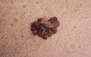
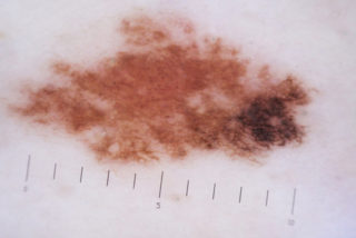
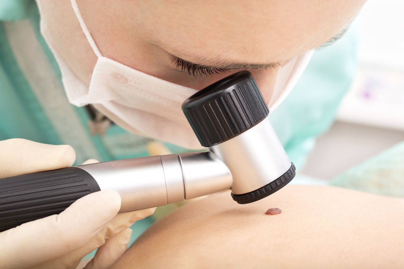
Despite the high rate of skin cancer in Australia, there are steps that people can take to protect themselves. The most important thing is to limit sun exposure, especially during peak hours when the sun’s rays are the strongest. This can be achieved by seeking shade, wearing protective clothing, and applying sunscreen with a high SPF. It is also important to learn how to detect skin cancer early and seek medical attention if they notice any changes in their skin. Early detection is key when it comes to treating skin cancer, and regular skin checks can help to identify any potential issues before they become more serious.
Australia’s high rate of skin cancer is a complex issue with several contributing factors. While the country’s location and outdoor lifestyle play a role, cultural attitudes towards tanning and a lack of awareness about the risks of sun exposure are also significant factors. It is important for Australians to take steps to protect themselves from the harmful effects of UV radiation, including limiting sun exposure, wearing protective clothing, and applying sunscreen. By taking these precautions, Australians can reduce their risk of developing skin cancer and enjoy the country’s beautiful outdoor lifestyle safely.
Need Help?
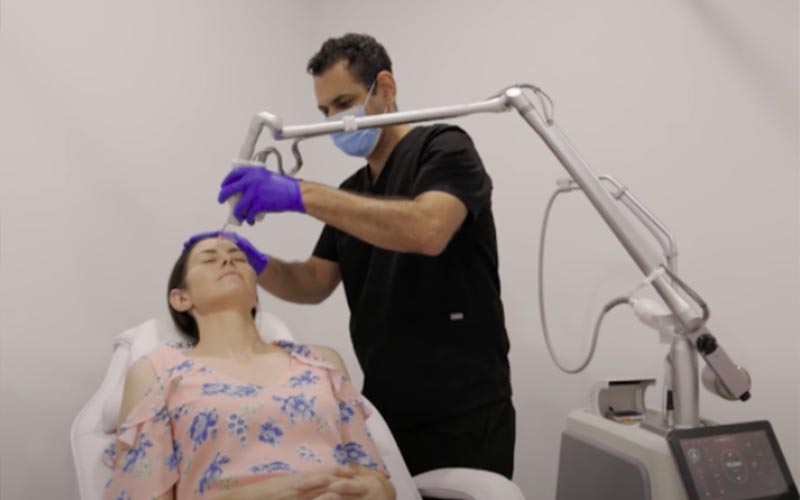
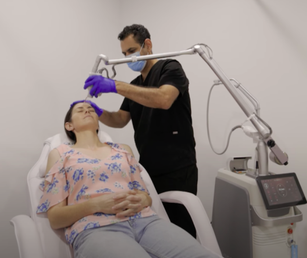
Elixir @ Hunter is a skin cancer cancer clinic in Maitland with with two operating rooms, a dressing room and a modern laser and cosmetic room. We also provide cosmetic treatment. If you are located in Newcastle and needing skin cancer treatment then we are only a short drive away.
Our coverage area: Newcastle, Singleton, Rutherford, Branxton, Cessnock, Aberglasslyn, Lochinvar