
How to check for melanoma or dysplastic moles at home?
Melanoma is the third commonest invasive malignancy in Australia after breast and prostate cancer. Checking your skin can help you find melanoma early when it is highly treatable. Consult your doctor if you ever think you have a concerning skin lesion. Examine your body for early signs of melanoma. Melanoma can affect you anywhere on your skin – from your scalp, hands to the bottom of your feet. Even if the area gets only a little sun, it is possible for melanoma to develop there. The highest reported rates of cutaneous melanoma in the world are in Australia and New Zealand. Melanoma can occur in adults of any age but is very rare in children.
Elixir @ Hunter is a purpose-built skin cancer clinic Maitland. Book your consultation today!
The main risk factors for developing the most common type of melanoma (superficial spreading melanoma) include:
- Increasing age
- Past history of Melanoma
- Past history of Non Melanoma skin cancers
- Many moles
- Multiple (>5) abnormal moles (histologically dysplastic moles)
- A strong family history of melanoma with 2 or more first-degree relatives affected
- White/fair skin that burns easily
How to check your own skin?
1. A full-length mirror
Looking into a full-length mirror helps check your back and sides.
2. Underarms, forearms, and palms
Bend your elbows and look carefully at your forearms, underarms, and your palms.
3. Legs, toes and soles of your feet
Look at the backs of your legs, feet, the spaces between your toes, and the soles of your feet.
4. A hand mirror can help you check your neck, scalp, back and buttocks
A small mirror can be handy to check your own back, buttocks and the back of your neck.
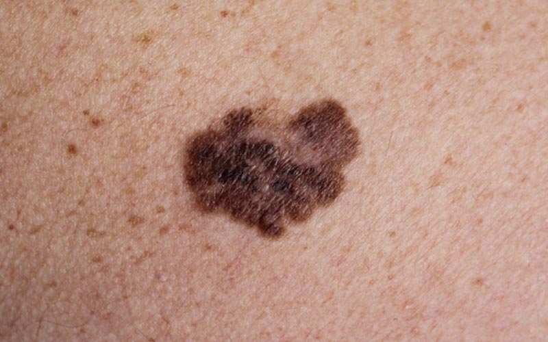
The ‘ABCDE’ approach to recognising an early melanoma
Remember the ABCDE when looking at each of your skin lesions.
A – Asymmetry
Melanomas are often neither circular nor oval in shape. If you draw a line through the middle of the lesion, the two halves do not match. This irregular shape is described as asymmetry.
B – Border
Melanoma borders tend to be uneven and may have scalloped or notched edges. Sometimes the edge of a melanoma is abrupt next to normal skin. At other times, the melanoma may merge into skin. These two border features can happen in different parts of the same melanoma.
C – Colour
Multiple colours are a warning sign. While benign moles are usually a single shade of brown, a melanoma may have different shades of brown, tan or black. As it grows, the colours red, white, or even blue may also appear. The more colours in the skin lesion, the more concerning it is.
D – Diameter
It is a warning sign if a skin lesion is 6 mm in diameter or greater. Most harmless skin lesions are quite small.
E – Evolution
Has your skin lesion changed over several months? This is a concerning feature. Harmless skin lesions often remain the same year after year. Inflamed skin lesions often change over days or weeks rather than over months.
Doctors generally do not use the ABCDE system
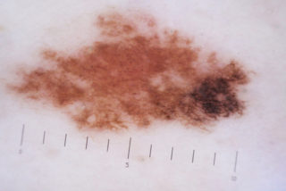
They usually examine your skin lesions with a dermatoscope. This is an instrument with magnification and a light source that reduces surface reflection. The doctor can detect the structure of the skin lesion under the surface, and use their knowledge and clinical skills to diagnose skin lesions suspicious for melanoma. The suspicious skin lesion will need removal or an excision biopsy. Alternatively, the dermatoscope may assist your doctor to determine the skin lesion is not concerning. The ABCDE system is just a guide, and it is not foolproof. A melanoma can be smaller than 5 mm in diameter. A melanoma can be circular and only have one colour.
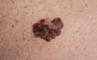
Dysplatic Naevi or Moles
A dysplastic melanocytic nevus is a mole with some structural abnormality. These dysplastic nevi are not skin cancers but patients with multiple dysplastic naevi are at a significant risk of developing melanoma.
Dysplastic moles look like very dark or black moles. They often have a strange irregular shape, are usually smooth to touch, and may rise gently off the skin. They are seen in people of every age. Other features may be ill defined or blurred borders, Irregular margin resulting in an unusual shape, varying shades of colour (mostly pink, tan, brown, black)
Dysplastic naevus syndrome is a condition where a patient has 5 or more dysplastic melanocytic naevi. These people are at substantial risk of developing a melanoma.
Patients with Dysplastic Naevus syndrome are at high risk of melanoma and so, It is therefore essential that extreme measures are taken to avoid sun damage. Of those people with dysplastic nevi who later develop melanoma, more grow from normal looking skin than from existing Dysplastic moles. As such, simply removing lots of dysplastic moles is not the answer. These patients need regular and careful skin checks to check for any changes. During skin checks, the doctor can take photographs of moles (with or without the dermatoscope) and any suspicious dysplastic moles and can then be followed up for any changes. The close-up photographs with dermatoscopic views should be repeated from time to time, so change can be detected early and its significance determined. Follow up is the key to managing dysplastic moles. If in doubt, a suspicious or changing atypical nevus should be removed for an excision biopsy. Partial biopsy is best avoided, as the test may miss a small focus of melanoma. People diagnosed with atypical naevi should be taught how to self examine their skin for new skin lesions and for changes to existing moles that may indicate the development of melanoma. People with numerous moles should have a thorough full body skin check 6-12 monthly.
Elixir @ Hunter offers skin & mole check in Newcaste, Maitland, Hunter Valley area.